PRODUCT 製品概要
ハイスループットスクリーニング
- 96ウェルプレート全面を約60秒で撮像、約30秒で解析 さらに、プレートスタッカーやインキュベータなど外部機器をプレート搬送ロボットで接続することで1日最大200枚の自動撮像が可能
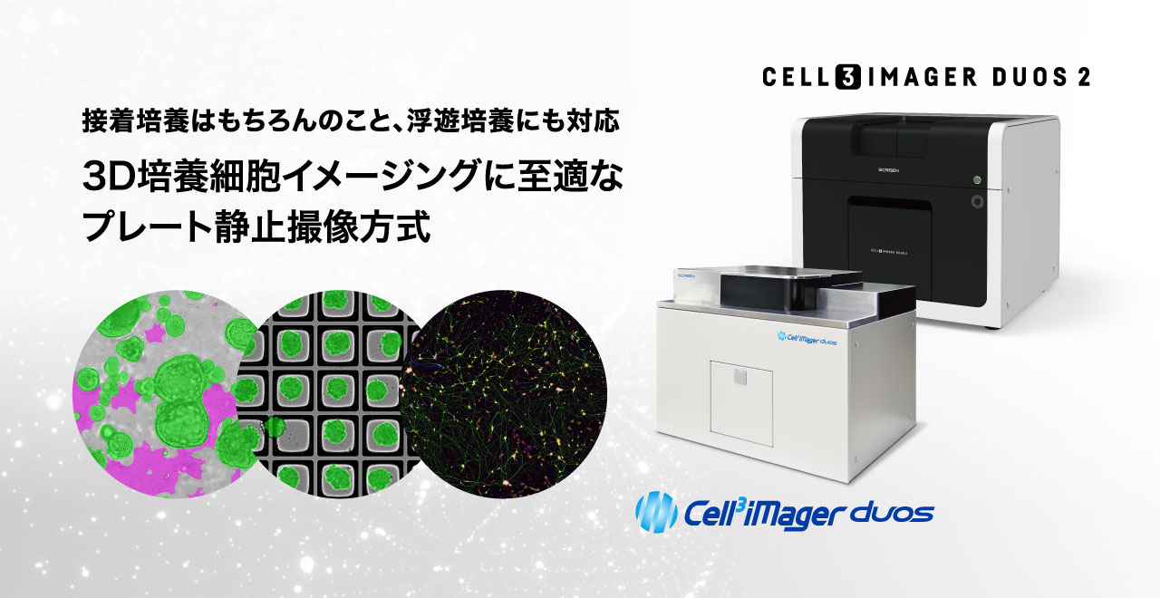
3次元培養細胞対応
- Zスタッキング撮像とフォーカス合成機能を搭載し、3次元培養細胞も解析可能。高さ方向に方向が異なる細胞もクリアに撮像
Deep Learning細胞抽出
- Deep Learning画像処理により、目的の細胞を"研究者の目"と同等に抽出。細胞形態ごとの抽出やハイコンフルエンシーかつ不均一な画像でも正確に定量可能
マルチカラー蛍光イメージング
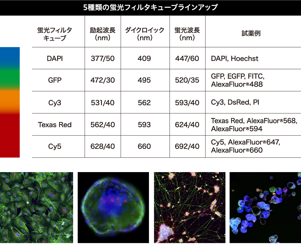
- LED蛍光フィルタキューブを搭載でき、明視野と蛍光色を自動で連続撮像、5種類の蛍光フィルタキューブをラインアップ
FEATURES 特徴
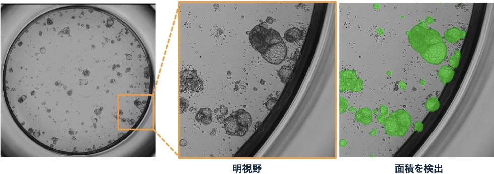
ホールウェルクリアスキャン
- メニスカスの影響を排除し、ウェル辺縁部をクリアに撮像
- ハイパーセントリック・テレセントリックの独自光学系の採用により、メニスカスの影響を低減してホールウェルを高画質に撮像し、ウェル辺縁部までの細胞を正確に計測
- 独自設計の解像度0.8um/pixと4um/pixのレンズを搭載
- 播種直後の細胞でも高精度に検出可能な細胞抽出アルゴリズムを搭載
- 高速モードは、96ウェル全面を60秒未満でスキャン可能
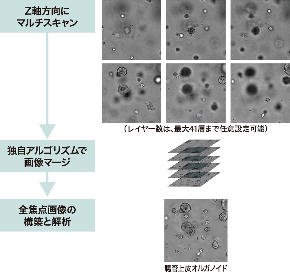
3次元培養細胞対応
- 3D培養細胞イメージングに至適な被写界深度が深い独自設計のレンズと照明を搭載
- Zスタッキング撮像とフォーカス合成機能を搭載
- F底、V底、U底の各種SBSフォーマット、マイクロウェルプレートに標準対応
- 3次元培養のためのプレート、Corning® Elplasia®(コーニングジャパン株式会社)、EZSPHERE(AGCテクノグラス株式会社)のような機能性特殊プレートにも対応
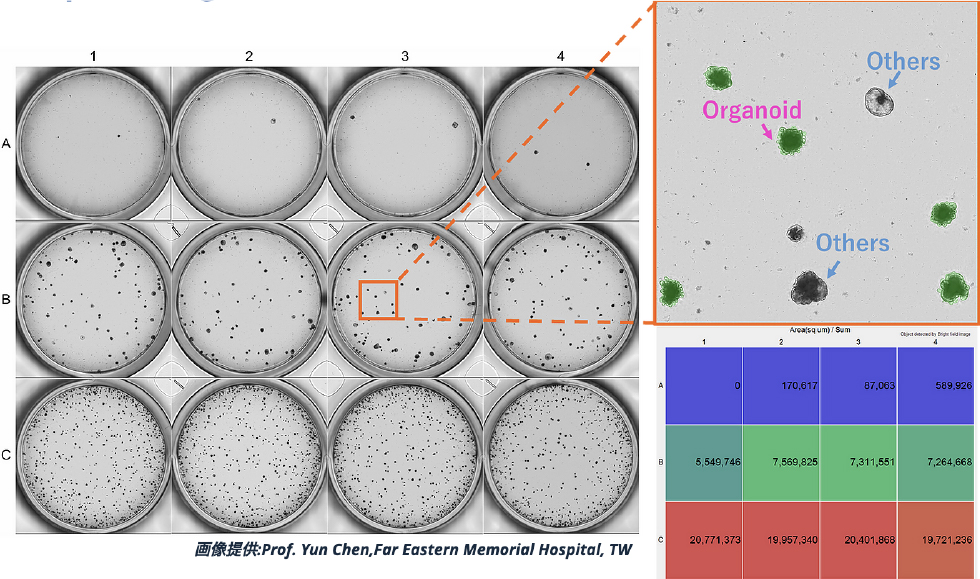
Deep Learning プラグイン (オプション)
- Deep Learningにより成長したオルガノイドのみを抽出、定量も可能
- ハイコンフルエンシーかつ不均一な画像もDeep Learningにより正確に抽出・計測可能
マルチカラー蛍光イメージング
- マルチカラー蛍光イメージングに対応
- LED蛍光フィルタキューブを3色まで同時に搭載でき※、明視野と蛍光3色を自動で連続撮像 ※ Cell3iMager duos2のみ
- 5種類の蛍光フィルタキューブをラインアップ
※ Cell3iMager duos2のみ
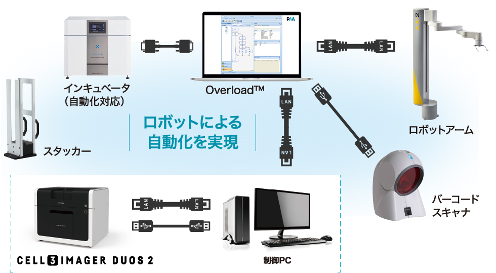
オートメーションシステム
- プレートスタッカーやインキュベータなど外部機器をプレート搬送ロボットで接続し自動化
- 1日最大200枚の自動撮像、煩雑なワークフローのロボット化により業務効率化
神経突起プラグイン (オプション)
- 神経突起の伸長計測など、マルチオブジェクト解析をプラグイン方式で提供
Publication 論文
-
Novel bone microenvironment model of castration‑resistant prostate cancer with chitosan fiber matrix and osteoblasts
Masahiro Samoto Hideyasu Matsuyama Hiroaki Matsumoto Hiroshi Hirata Koji Ueno Sho Ozawa Junichi Mori Ryo Inoue Seiji Yano Yoshiaki Yamamoto Jun Haginaka Shizuyo Horiyama Koji Tamada
August 1, 2021 https://doi.org/10.3892/ol.2021.12950 Oncology Letters Article Number: 689 -
Mammary cell gene expression atlas links epithelial cell remodeling events to breast carcinogenesis
Kohei Saeki, Gregory Chang, Noriko Kanaya, Xiwei Wu, Jinhui Wang, Lauren Bernal, Desiree Ha, Susan L. Neuhausen, and Shiuan Chen
Commun Biol. 2021; 4: 660. Published online 2021 Jun 2. doi:10.1038/s42003-021-02201-2 -
Lactate Reprograms Energy and Lipid Metabolism in Glucose-Deprived Oxidative Glioma Stem Cells
Noriaki Minami ORCID,Kazuhiro Tanaka ,Takashi Sasayama ,Eiji Kohmura ORCID,Hideyuki Saya andOltea Sampetrean
Metabolites 2021, 11(5), 325; https://doi.org/10.3390/metabo11050325 Published: 18 May 2021
APPLICATION アプリケーション
-
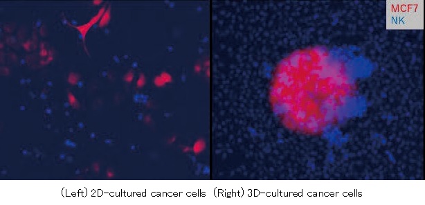
2D/3D培養がん細胞に対する NK細胞 Killing Assay
近年、創薬開発やがん研究分野においてNK(Natural Killer)細胞が注目されています。NK細胞は自然免疫で働くリンパ球の一種であり、特に腫瘍細胞やウイルス感染細胞の除去に主要な役割を果たしています。Cell3iMager duos / duos2およびDeep Learningプラグインを使用した明視野解析により、長期間のKilling Assayを行うことができるようになります。
-
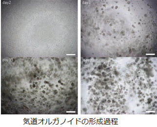
ヒト iPS 細胞由来 気道オルガノイドを用いた 疾患モデリングとイメージングアッセイ
呼吸器疾患は、慢性進行性の疾患や、昨今のCOVID-19肺炎などの急性かつ重症の疾患など、幅広い病態が存在する。しかし、本領域における根本的な治療薬の開発は遅れており、その原因の一つとして、前臨床の薬効評価などに使われるマウスなどの動物モデルが、ヒトの病態を反映しない場合が多いことが挙げられる。その代表例として、塩素イオンチャネルであるcystic fibrosis transmembrane conductance regulator (CFTR)の変異による常染色体劣性遺伝性疾患である嚢胞性線維症(Cysticfibrosis,CF)が挙げられる。
近年、オルガノイドと呼ばれる臓器様三次元構造を体外で構築し、疾患モデリングや創薬等に応用することが出来るようになってきた。その中で、嚢胞性線維症患者より樹立されたiPS細胞から分化誘導した気道上皮オルガノイドを用いて、フォルスコリンアッセイを行い、そのフェノタイプをラベルフリーイメージングで捉えることができた。本実験で用いた評価系は、Cell3iMager duosでの撮像および画像データ取得を行い、ディープラーニングでオルガノイド領域をセグメンテーションし、面積を計測することでオルガノイドの膨張率を定量した。
ラベルフリーのため手間も少なく、かつディープラーニングに基づく客観的な計測により、正確性、汎用性そして高スループット性があるため、上記ニーズに応えて嚢胞性線維症治療の進展に寄与することが期待される。
資料請求はこちら -
2D/3D培養がん細胞に対する NK細胞 Killing Assay
近年、創薬開発やがん研究分野においてNK(Natural Killer)細胞が注目されています。NK細胞は自然免疫で働くリンパ球の一種であり、特に腫瘍細胞やウイルス感染細胞の除去に主要な役割を果たしています。Cell3iMager duos / duos2およびDeep Learningプラグインを使用した明視野解析により、長期間のKilling Assayを行うことができるようになります。
-
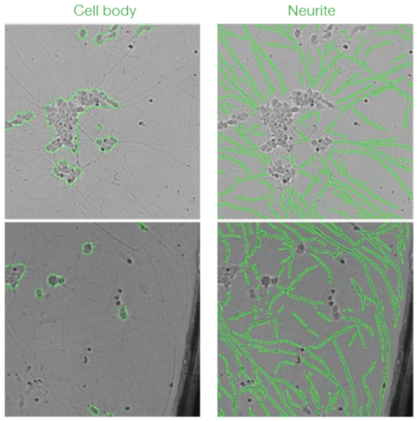
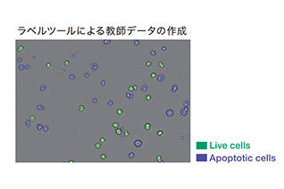
Cell3iMager duos を用いたiPS 細胞由来神経細胞の明視野解析
Cell3iMager duosおよびディープラーニングを用いてヒトiPS細胞由来交感神経細胞の明視野解析ができることを確認しました。従来の画像処理方法に比べてロバスト性が高く、明るさの違いがあったり、デブリの存在下でも、細胞を正確に抽出することが可能です。また、アポトーシスが誘導された細胞も抽出することが可能なため、培養を続けながら生細胞と死細胞を定量的に評価することが可能となります。このことから、細胞の増殖性や品質の評価をラベルフリーで行うことが可能であり、例えば、神経細胞への長期的な薬剤暴露試験などに応用できる可能性があります。
-
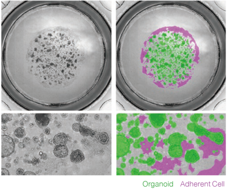
Cell3iMager duos / duos2 を用いたオルガノイドのラベルフリー解析
Cell3iMager duos / duos2を用いることで三次元培養したオルガノイドの明視野/蛍光イメージングが可能になります。また、ディープラーニングを用いることで、オルガノイド形状毎のセグメンテーションが可能であるため、これまで顕微鏡観察で行ってきたオルガノイド成長過程のモニタリングをラベルフリー解析で代替できる可能性があります。同様に、ATPアッセイや蛍光染色で行ってきた毒性評価、FISアッセイなどをラベルフリー解析で代替できる可能性があります。
-
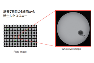
Single cell cloning using Hybridoma
ハイブリドーマ4B2細胞の1細胞クローニングをCell3iMager duosを用いて行った。
-
Single Cell Cloning
HCT116細胞の1細胞クローニングをCell3iMager duosを用いて行った。
-
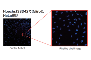
Counting fluorescence stained adherent cells
単層培養した細胞を蛍光染色することによって細胞数の計測を行った。
-
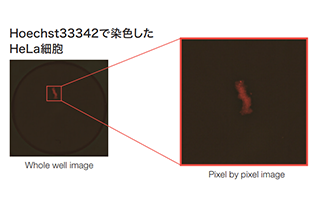
Evaluation of hypoxia level in 2D/3D-cultured cells
単層または三次元培養時におけるHypoxiaレベルをHypoxia probeによる蛍光染色で定量比較した。
-
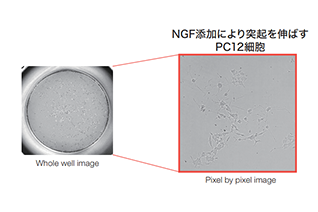
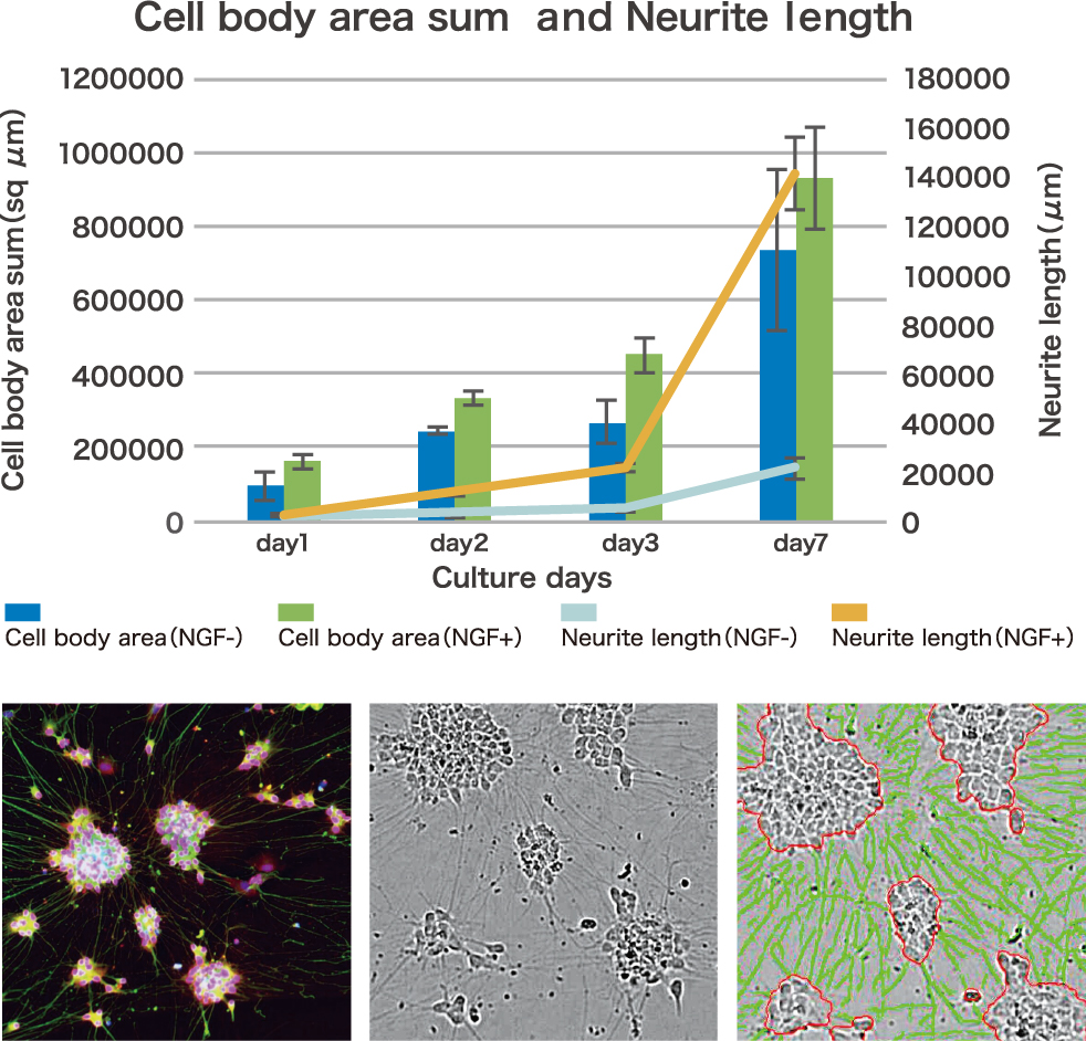
Neuronal Differentiation
神経成長因子(β-NGF)を添加したPC12細胞の分化効率をCell3iMager duosを用いてラベルフリーで定量した。
-
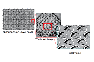
Analysis of spheroids in microwells (EZSPHERE® plate)
EZSPHERE®プレートでスフェロイドを作製し、ディープラーニング機能によりラベルフリー解析を行った。
-
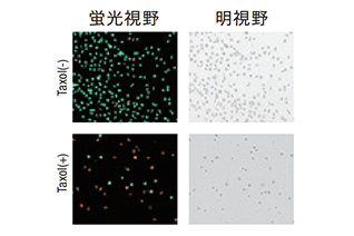
Live/Dead Assay
抗がん剤(Taxol)処理したU937細胞の生存率をCalcein-AMとPIによる蛍光撮像により解析した。
-
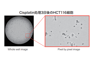
Cell Viability and Cytotoxicity Assay
抗がん剤(Cisplatin)処理したがん細胞の生存率をCell3iMager duosを用いて ラベルフリーで定量した。
-
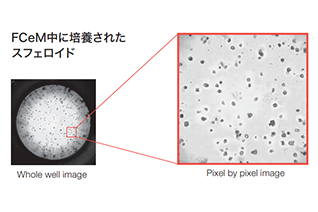
3D Cell Culture by using FCeM®
FCeM培地中で培養されたスフェロイドをCell3iMager duosを用いて ラベルフリーで定量した。
-
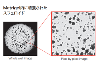
Assessing the growth of 3D spheroids cultured in Matrigel®
段階的な細胞数で細胞を播種し、Cell3iMager duosを用いて細胞密度を定量化した。
-
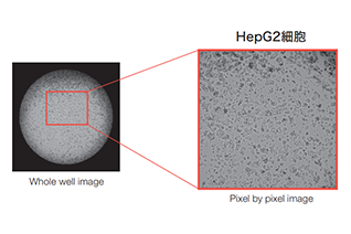
Confluency
段階的な細胞数で細胞を播種し、Cell3iMager duosを用いて 細胞密度を定量化した。
-
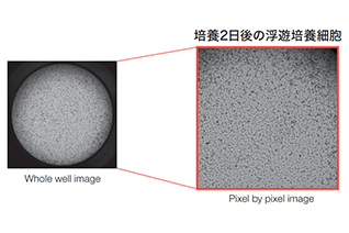
Suspension cultured cells
浮遊培養細胞数をCell3iMager duosを用いてラベルフリーで定量した。
-
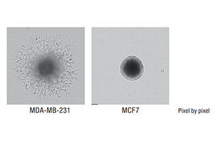
3D Invasion Assay
Cell3iMager duosを用いて、悪性度の異なる乳がん細胞の浸潤能を比較した。
-
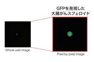
Anti-Cancer Drug Screening
Cell3iMager duosを用いて、3次元培養下TCF4/LEFレポーター活性をGFP蛍光強度で定量化し、薬剤スクリーニングを行った。その結果、Wntシグナル阻害活性を示す化合物Sが大腸がん治療薬の候補として同定された。
-
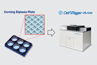
Label free analysis of spheroids in microwells (Corning® Elplasia® Plates)
CORNING社のマイクロウェルプレートCorning®Elplasia®で培養したスフェロイドをCell3iMager duosを用いて撮像・解析を行った。
-
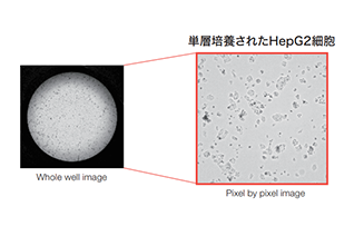
Cell Proliferation
単層/三次元培養された細胞の増殖率をCell3iMager duosを用いてラベルフリーで定量した。
-
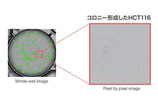
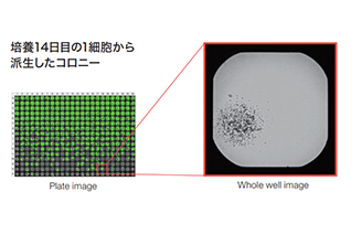
Label free colony-forming units assay
がん細胞をCell3iMager duosを用いてラベルフリーでコロニーフォーメーションアッセイ(CFUアッセイ)を行った。
-
Deep Learning for cell classification
Anti-Fas抗体により細胞死を誘導したJarkat細胞についてCell3iMager duosとDeep Learingを用いて形態分類を行った
Publication 論文
-
Scalable production of homogeneous cardiac organoids derived from human pluripotent stem cells
Taijun Moriwaki Hidenori Tani Kotaro Haga Yuika Morita-Umei Yusuke Soma Tomohiko C. Umei Otoya Sekine Kaworu Takatsuna Yoshikazu Kishino Hideaki Kanazawa Jun Fujita Keiichi Fukuda Shugo Tohyama Masaki Ieda
-
Artificial intelligence-based analysis of time-lapse images of sphere formation and process of plate adhesion and spread of pancreatic cancer cells
Yuuki Shichi, Fujiya Gomi1, Yasuko Hasegawa, Keisuke Nonaka, Seiichi Shinji, Kimimasa Takahashi, Toshiyuki Ishiwata
-
Novel and efficient method for culturing patient‐derived gastric cancer stem cells
Tomonori Morimoto, Yukitoshi Takemura,Takemitsu Miura,Takehito Yamamoto,Fumihiko Kakizaki,Hideo An,Hisatsugu Maekawa,Tadayoshi Yamaura, ,Kenji Kawada, Yoshiharu Sakai,Yoshiaki Yuba,Hiroaki Terajima,Kazutaka Obama,Makoto Mark Taketo,and Hiroyuki Miyoshi
-
Suspension culture in a rotating bioreactor for efficient generation of human intestinal organoids
Junichi Takahashi Tomohiro Mizutani HadyYuki Sugihara Sei Kakinuma Mamoru Watanabe Ryuichi Okamoto
-
Derivation of pancreatic acinar cell carcinoma cell line HS-1 as a patient-derived tumor organoid
Daisuke Hoshi,Emiri Kita,Yoshiaki Maru,Hiroyuki Kogashi,Yuki Nakamura,Yasutoshi Tatsumi,Osamu Shimozato,Kazuyoshi Nakamura,Kentaro Sudo,Akiko Tsujimoto,Ryo Yokoyama,Atsushi Kato,Tetsuo Ushiku,Masashi Fukayama,Makiko Itami,Taketo Yamaguchi,Yoshitaka Hippo
SPECIFICATIONS スペック
| 品名(型式) | Cell3iMager duos2 (CC-8300) | Cell3iMager duos (CC-8000) |
|---|---|---|
| チャンネル | 明視野 + 蛍光3色搭載 | 明視野 + 蛍光1色搭載 |
| 明視野光源 | ストロボ白色LED | ストロボ白色LED |
| カメラ | CMOS 4.2メガピクセル カラー | CMOS 4.2メガピクセル カラー |
| 光学系 | 独自ハイパーセントリック光学系 (高速モード時) 独自テレセントリック光学系 (高精細モード時) |
独自ハイパーセントリック光学系 (高速モード時) 独自テレセントリック光学系 (高精細モード時) |
| 分解能 | 4.0um/pixel相当 (高速モード時) 0.8um/pixel相当 (高精細モード時) |
4.0um/pixel相当 (高速モード時) 0.8um/pixel相当 (高精細モード時) |
| オートフォーカス | HW: レーザー式リアルタイムオートフォーカス SW: 画像コントラストソフトウエアオートフォーカス |
HW: レーザー式リアルタイムオートフォーカス SW: 画像コントラストソフトウエアオートフォーカス |
| 画像出力 | 24bit color (8bit×3) | 24bit color (8bit×3) |
| 蛍光フィルタキューブ | DAPI, GFP, Cy3, Texas Red, Cy5 | ‐ |
| 蛍光光源(励起波長) | ‐ | U : 384nm , B : 470nm , G : 530nm , Y565nm , R : 625nm |
| 庫内温度 | 35℃±2℃に自動調整(装置電源On時) | 35℃±2℃に自動調整(装置電源On時)※オプション |
| 設置環境 | 室温18‐28℃, 湿度80%以下(結露なきこと) | 室温18‐28℃, 湿度80%以下(結露なきこと) |
| 輸送条件 | 梱包状態で0‐55℃, 湿度80%以下(結露なきこと) | 梱包状態で0‐55℃, 湿度80%以下(結露なきこと) |
| 培養容器 | 6, 12, 24, 48, 96, 384 wellプレート(ほとんど全てのSBS規格適合品に対応) 35, 60,100mm ディッシュ / スライドグラス(別途アダプタが必要) |
6, 12, 24, 48, 96, 384 wellプレート(ほとんど全てのSBS規格適合品に対応) 35, 60,100mm ディッシュ / スライドグラス(別途アダプタが必要) |
| 電源 | AC100-240V / 250VA | AC100-240V / 250VA |
| 外寸・重量 | W677 x D580 x H550 mm / 111kg | W677 x D570 x H550 mm / 110kg |
| ソフトウエア | Cell3iMager専用ソフトウエア(標準付属) | Cell3iMager専用ソフトウエア(標準付属) |
| 指定コンピュータ(動作確認済) | HP Z4 G4 ワークステーション(弊社指定構成), OS: Windows 10 | HP Z4 G4 ワークステーション(弊社指定構成), OS: Windows 10 |
| 品名(型式) | チャンネル | 明視野光源 | カメラ | 光学系 | 分解能 | オートフォーカス | 画像出力 | 蛍光フィルタキューブ | 蛍光光源(励起波長) | 庫内温度 | 設置環境 | 輸送条件 | 培養容器 | 電源 | 外寸・重量 | ソフトウエア | 指定コンピュータ(動作確認済) |
|---|---|---|---|---|---|---|---|---|---|---|---|---|---|---|---|---|---|
| Cell3iMager duos2 (CC-8300) |
明視野 + 蛍光1色搭載 | ストロボ白色LED | CMOS 4.2メガピクセル カラー | 独自低被写界深度光学系 | 4.0um/pixel相当 (高速モード時) / 0.8um/pixel相当 (高精細モード時) |
HW: レーザー式リアルタイムオートフォーカス SW: 画像コントラストソフトウエアオートフォーカス |
24bit color (8bit×3) | DAPI, GFP, Cy3, Texas Red, Cy5 | - | 35℃±2℃に自動調整(装置電源On時) | 室温18‐28℃, 湿度80%以下(結露なきこと) | 梱包状態で0‐55℃, 湿度80%以下 (結露なきこと) |
6, 12, 24, 48, 96, 384 wellプレート (ほとんど全てのSBS規格適合品に対応) 35, 60,100mm ディッシュ スライドグラス (別途アダプタが必要) |
AC100-240V / 250VA | W677 x D580 x H550 mm / 111kg | Cell3iMager専用ソフトウエア(標準付属) | HP Z4 G4 ワークステーション(弊社指定構成), OS: Windows 10 |
| Cell3iMager duos (CC-8000) |
- | U : 384nm , B : 470nm , G : 530nm , Y565nm , R : 625nm |
35℃±2℃に自動調整(装置電源On時) ※オプション |
W677 x D570 x H550 mm / 110kg |