Information
2022.01.01
The 22nd Annual Meeting of the Japanese Society for Regenerative Medicine
Chairman: Jun Takahashi (Director, Center for iPS Cell Research and Application, Kyoto University)
Date: March 23 (Thu) to 25 (Sat), 2023
Place: Kyoto International Conference Center
Clear bright-field image acquisition by high-definition stitching technology
By using high-definition stitching technology based on our proprietary image processing and image analysis technologies, clear bright-field images can be obtained even at high magnifications.
It is especially suitable for applications such as cloning, which requires accurate cell counting.
SCREEN
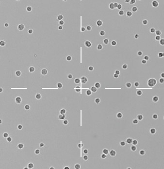
High-precision image processing technology makes it possible to measure accurately with virtually no discernible image seams.
Conventional
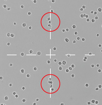
Misalignment may occur at image joints, affecting accurate cell counts.
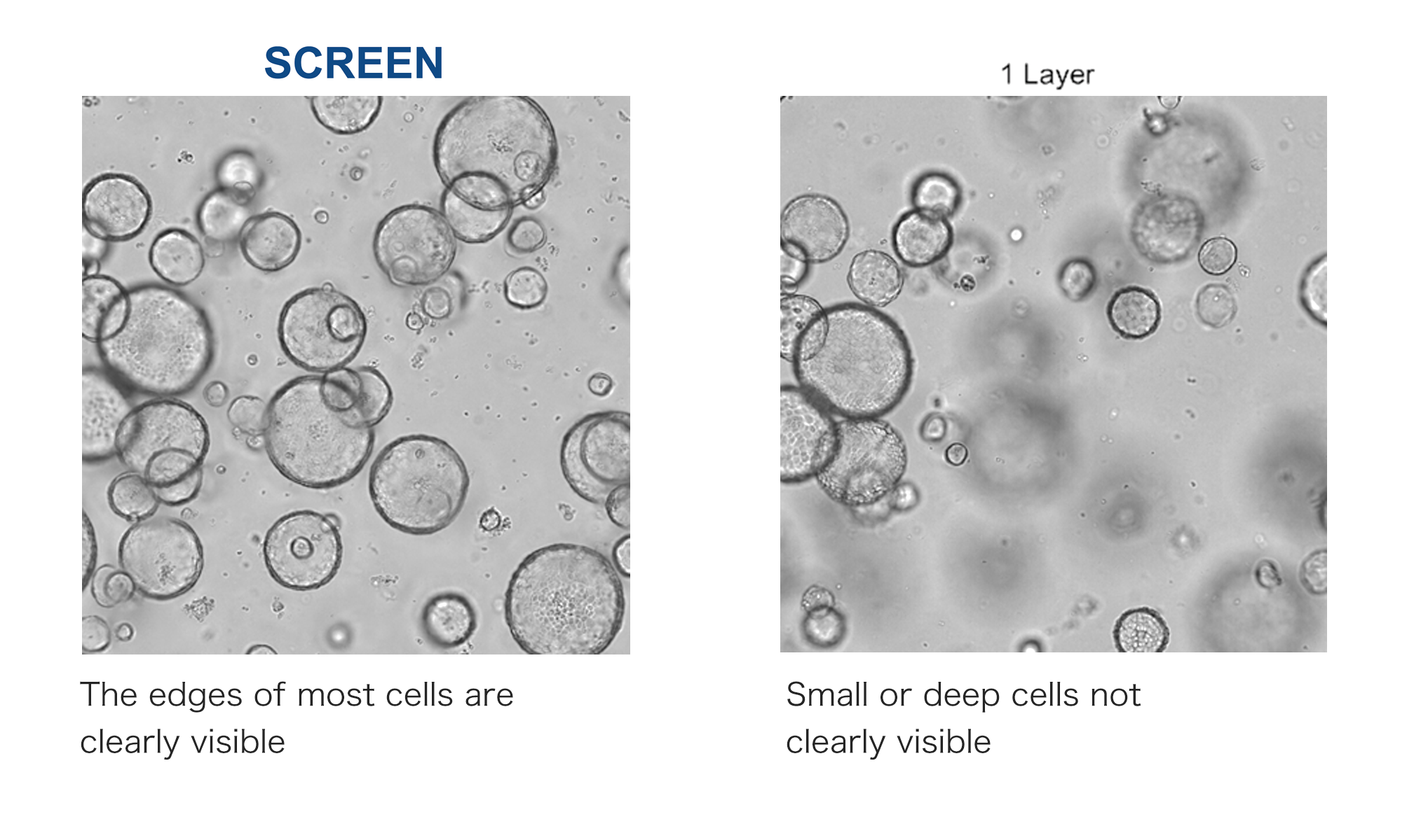
All-in-focus images optimal for 3D cultured cell analysis can be acquired
Multiple images are captured while varying the focus in the Z direction and combined into a single image Supports various SBS plate formats such as F-bottom, V-bottom, U-bottom, etc.
Designed a simple and intuitive GUI design
Equipped with security-related functions
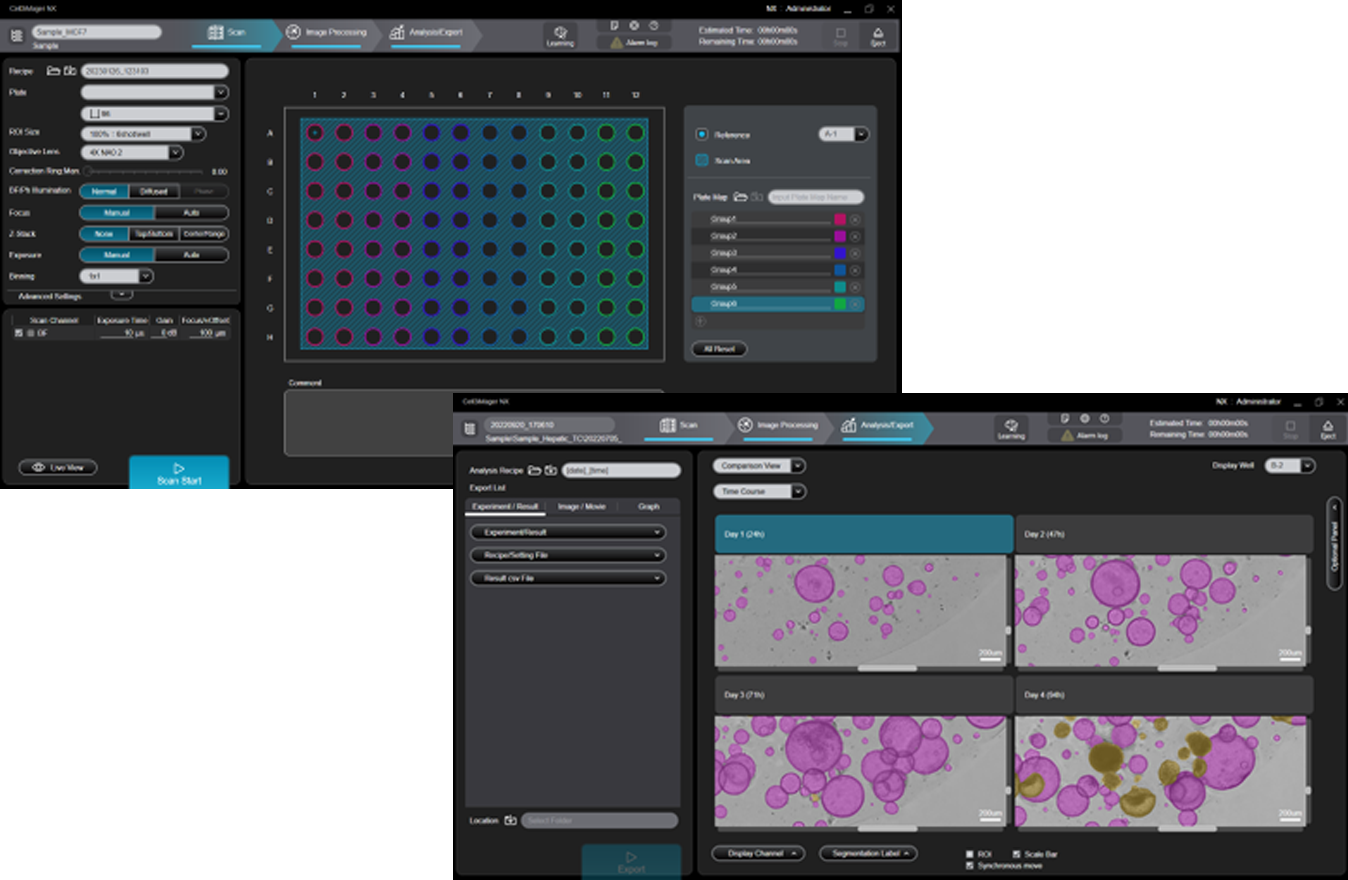
Imaging, measurement, and analysis can be performed on a single software package.
Useful security management functions such as database management, audit trails, and operation authority management.
Data can be output in various formats such as heat maps and scatter plots.
OPTION
Deep Learning Plug In
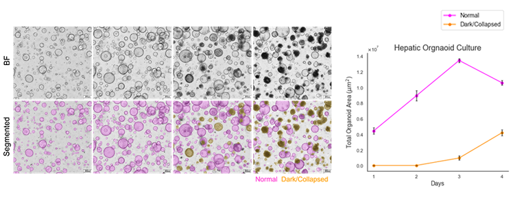
Deep Learning enables extraction and quantification of only grown organoids.
Deep Learning enables accurate extraction and measurement of high-confluency and non-uniform images.
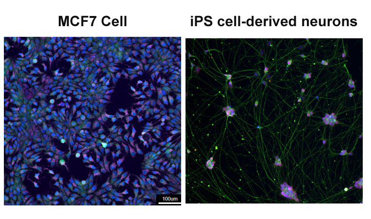
Support for multi-fluorescence analysis
Supports multi-color fluorescence imaging.
Up to 5 fluorescent filters can be installed simultaneously, allowing automatic continuous imaging of brightfield and 4 fluorescent colors.
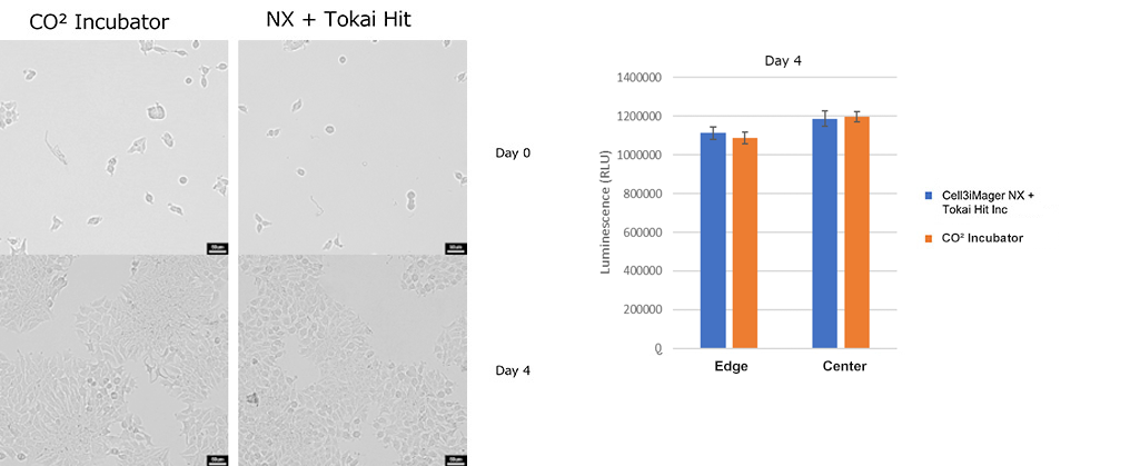
Can be equipped with a culture system for microscope from Tokai Hit Co. This system contributes greatly to work efficiency in cell culture monitoring.
APPLICATION
Ideal plate static imaging method for 3D cultured cell imaging
Specifications
| Product Name(Code) | Cell3iMager NX (CC-100) |
| Lighting unit | Strobe White LED / Aperture stop (2 types), phase difference, automatic shutter switching |
| Camera | CMOS 12M pixel monochrome |
| Auto Focus | Light source Laser diode / Detection range -0.5mm to 3.5mm |
| Magnification | Standard / 4x, 10x Objective lens Option / 2x, 20x, 40x Objective lens, 10x, 20x phase difference |
| Supported Plate | 6,12,24,48,96,384 well plate / 35,60,100 mm dish |
| Out put | 8bit mono |
| Channel | Bright field and 4 Colors of Fluorescence |
| Power Supply | 100–240VAC / 190VA |
| Size / Weight | W500 × D500 × H530 mm, 44 kg |
| Internal temperature | 18℃ ± 2℃ ~40℃ ± 2℃ Adjustment in 1℃ increments (without cooling mechanism) |
| Environment | RT 18-28℃, Humidity 80% or Less, no condensation |
| Software | Cell3iMager NX Specialized software(Standard included) |
| Control Computer | DELL Precision 3660 Tower / DELL monitor E2222H (Operation has been verified with our specified configuration) |