- APPLICATION
Drug discovery
-
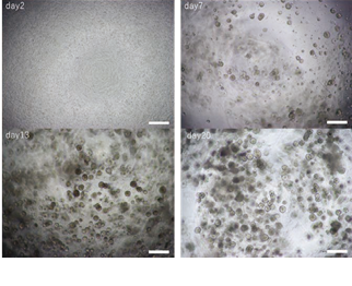
Disease modeling and imaging assays using human iPS cell-derived airway organoids
Respiratory diseases include a wide range of conditions, including
chronic progressive diseases and acute and severe diseases such as
the recent COVID-19 pneumonia. However, the development of
fundamental therapeutics in this field has been slow, partly because
animal models such as mice used for preclinical drug efficacy evaluation
often do not reflect human disease states. A typical example is
cystic fibrosis (CF), an autosomal recessive genetic disorder caused
by mutations in the cystic fibrosis transmembrane conductance
regulator (CFTR), a chloride ion channel. -
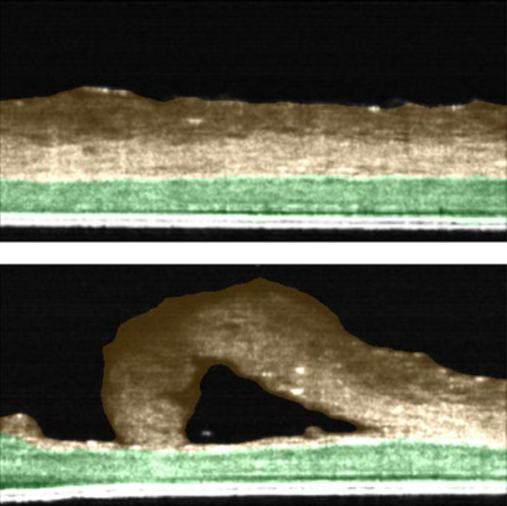
Skin Irritation test : Evaluation of thickness in skin model using OCT technology
To summarize, we observed that by using the low magnification mode, we were able to obtain the thickness information of the entire 24-well insert. In high magnification mode, we were able to obtain thickness information of two layers, stratum corneum and cell layer, non-invasively. We conclude that the Cell3iMager Estier equipped with OCT technology can be used to measure the thickness of stacked cell sheets in real time and implemented as “Skin irritation test” to evaluate safety of topical creams/agents developed by Biotech and Pharma industry.
-
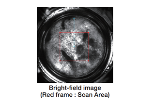
Thickness of cell sheet
NHDF sheets were imaged by Cell3iMager Estier, and thickness of sheets were calculated from these images.
-
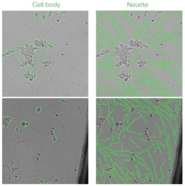
Imaging and Deep Learning based analysis of neurite outgrowth
With this study we conclude that Cell3iMager duos imaging technology in conjunction with deep learning is highly suited for delineating various biological processes using sympathetic neurons derived from hiPSC. Additionally, the platform can be used for identifying drug entities that could stimulate or block neuronal outgrowth. The analysis by deep learning accurately detects cells and is highly robust compared to traditional analyses even if there are differences in brightness or in the presence of debris.
-
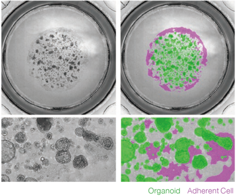
Label free imaging and analysis of organoids using Cell3iMager
Cell3iMager duos / duos2 technology is highly suited for brightfield/fluorescence imaging of 3D cultured organoids. We conclude that the Label-free analysis is highly advantageous and could substitute microscopic method of monitoring of organoid growth processes, as segmentation of each organoid shape is possible using Deep Learning. Similarly, label-free analysis may also substitute ATP assays, toxicity assessments performed by fluorescent staining, and FIS assays.
-
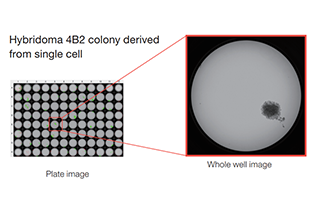
Single cell cloning using Hybridoma
Isolation of single cell derived clone with Duos superior imaging technology.
-
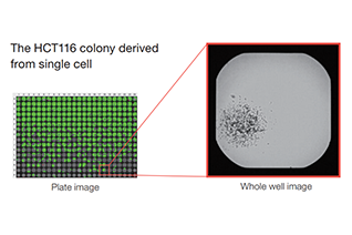
Single Cell Cloning
Utility of Cell3iMager duos imaging platform for isolation and assessment of single cell derived clone
-
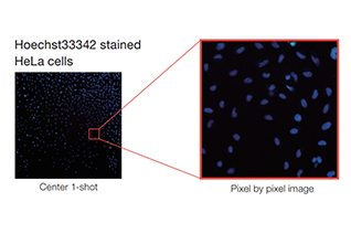
Counting fluorescence stained adherent cells
The cell density that stepwise cell number seeding, was quantyfied by using Cell3iMager duos.
-
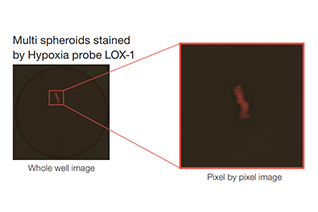
Evaluation of hypoxia level in 2D/3D-cultured cells
Hypoxia level of 2D/3D-cultured cells was evaluated by using Cell3iMager duos.
-
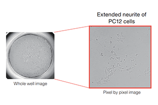
Neuronal Differentiation
Differentiation efficiency of β-NGF-treated PC12 was evaluated by using Cell3iMager duos.
-
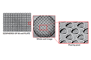
Analysis of spheroids in microwells (EZSPHERE® plate)
Spheroids were cultured with EZSPHERE® PLATE and analyzed using Deep Learning plug-in.
-
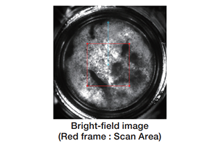
Thickness of cell sheet
NHDF sheets were imaged by Cell3iMager Estier, and thickness of sheets were calculated from these images.
-
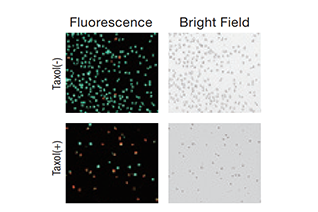
Live/Dead Assay
Cell viability in the presence of drug is calculated by using Calcein-AM/PI-double staining.