- APPLICATION
Regenerative medicine
-
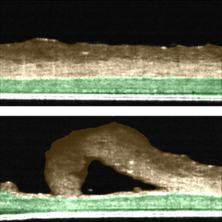
Skin Irritation test : Evaluation of thickness in skin model using OCT technology
To summarize, we observed that by using the low magnification mode, we were able to obtain the thickness information of the entire 24-well insert. In high magnification mode, we were able to obtain thickness information of two layers, stratum corneum and cell layer, non-invasively. We conclude that the Cell3iMager Estier equipped with OCT technology can be used to measure the thickness of stacked cell sheets in real time and implemented as “Skin irritation test” to evaluate safety of topical creams/agents developed by Biotech and Pharma industry.
-
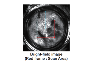
Thickness of cell sheet
NHDF sheets were imaged by Cell3iMager Estier, and thickness of sheets were calculated from these images.
-
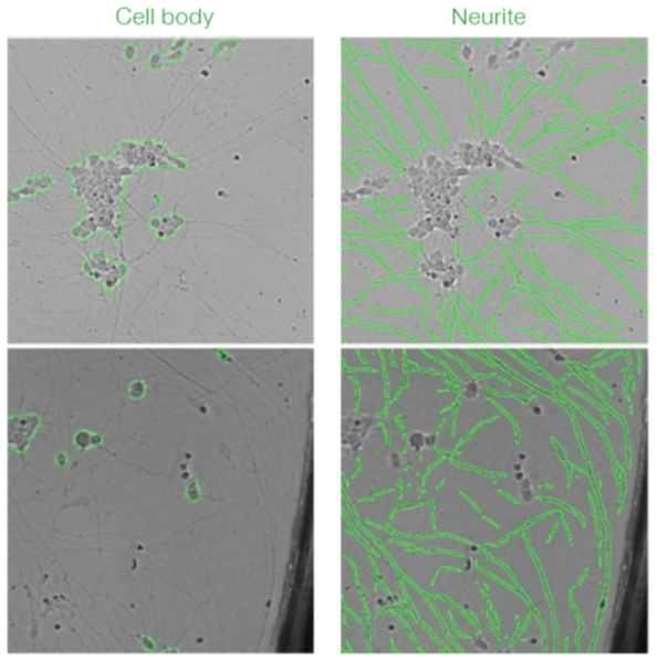
Imaging and Deep Learning based analysis of neurite outgrowth
With this study we conclude that Cell3iMager duos imaging technology in conjunction with deep learning is highly suited for delineating various biological processes using sympathetic neurons derived from hiPSC. Additionally, the platform can be used for identifying drug entities that could stimulate or block neuronal outgrowth. The analysis by deep learning accurately detects cells and is highly robust compared to traditional analyses even if there are differences in brightness or in the presence of debris.
-
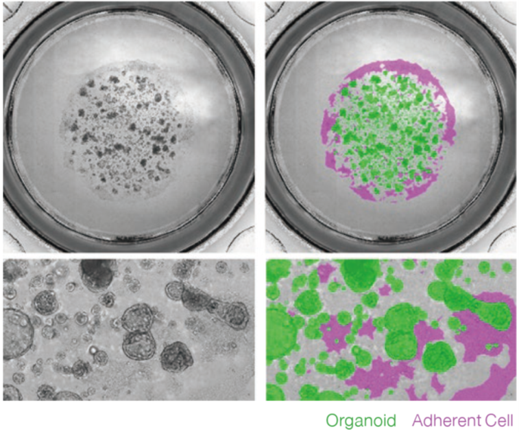
Label free imaging and analysis of organoids using Cell3iMager
Cell3iMager duos / duos2 technology is highly suited for brightfield/fluorescence imaging of 3D cultured organoids. We conclude that the Label-free analysis is highly advantageous and could substitute microscopic method of monitoring of organoid growth processes, as segmentation of each organoid shape is possible using Deep Learning. Similarly, label-free analysis may also substitute ATP assays, toxicity assessments performed by fluorescent staining, and FIS assays.
-
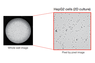
Cell Proliferation
Proliferation ratio of the 2D/3D cultured cells is quantified by Cell3iMager duos.
-
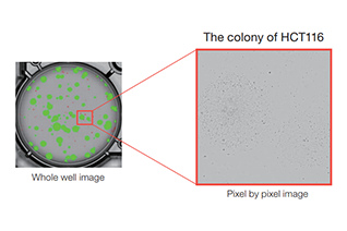
Label free colony-forming units assay
We performed the labeled-free colony formation assay using Cell3iMager duos.
-
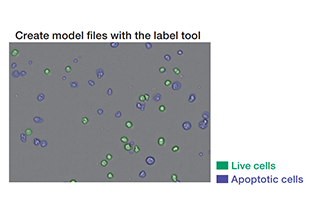
Deep Learning for cell classification
Jarkat cells induced cell death by Anti-Fas antibody were classified by Cell3iMager duos and Deep Learning.
-
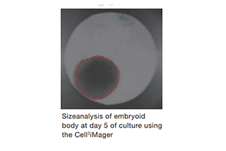
Rapid production and size assessment of Embryoid Bodies ( EBs )
Simplify formation of EBs with GravityPLUS™ hanging drop plates and Cell3 iMager analysis
-
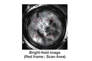
Thickness of cell sheet
NHDF sheets were imaged by Cell3iMager Estier, and thickness of sheets were calculated from these images.
-
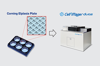
Label free analysis of spheroids in microwells (Corning® Elplasia® Plates)
Spheroids in microwells of Corning®Elplasia® plate made by Corning are scanned and measured by Cell3iMager duos.
-
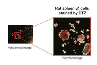
Pancreatic β cells stained by DTZ
Pancreatic β cells isolated from Rat spleen was quantified by Cell3iMager neo. To detect only β cells, isolated cells were stained by DTZ.
-

Label free colony-forming units assay
We performed the labeled-free colony formation assay using Cell3iMager duos.