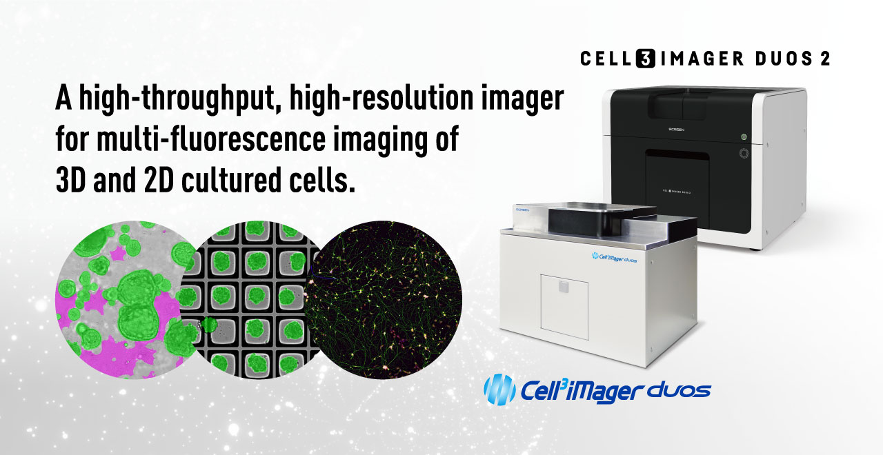

PRODUCT
High throughput
- 96-well plates can be imaged in about 60 seconds and analyzed in about 30 seconds.
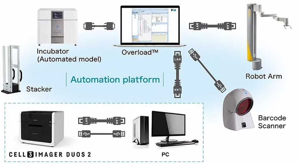
- In addition, automatic capture of up to 200 plates per day is possible by connecting external devices such as plate stackers and incubators with a plate transfer robot.
3D cultured cells
- 3D cultured cells can be analyzed with Z-stacking imaging and focus composition functions. Even cells scattered in the height direction can be captured clearly.
Deep Learning Cell Segmentation
- Deep learning image processing enables "researcher's eye" segmentation of target cells. Accurate quantification is possible even for segmentation by cell morphology and high confluency, non-uniform images.
Multi-fluorescence imaging
- Fluorescent color and bright field are automatically and continuously captured.
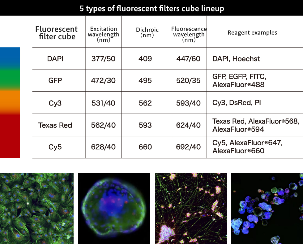
- 5 types of fluorescent filters are available.
FEATURES
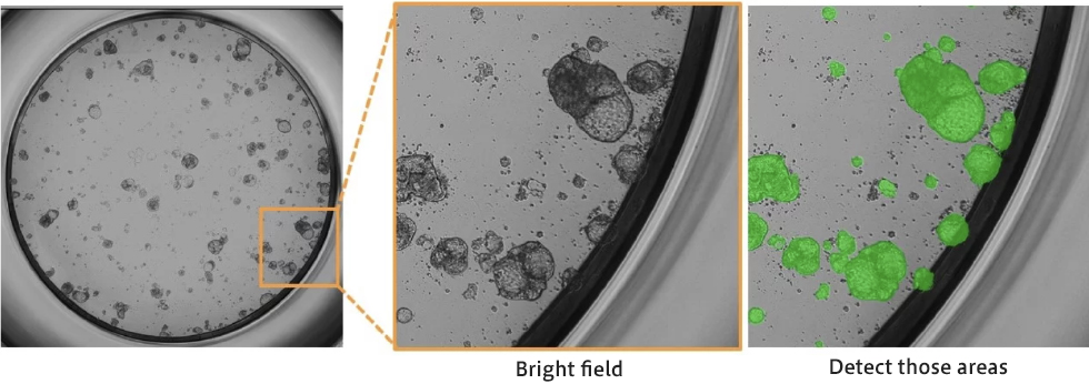
Meniscus-less
- Less meniscus and clear imaging even at the peripheral area
- Unique hyper-centric and tele-centric optical systems enables
a high-resolution imaging of whole well including the marginal area of well - Lens have 2 resolution which has 0.8um/pix and 4um/pix
- Equipped with high accurate extraction algorithm even cells soon after seeing
- High speed mode provide 60 sec/ 96 well plate and 70 sec/ 384 well plate
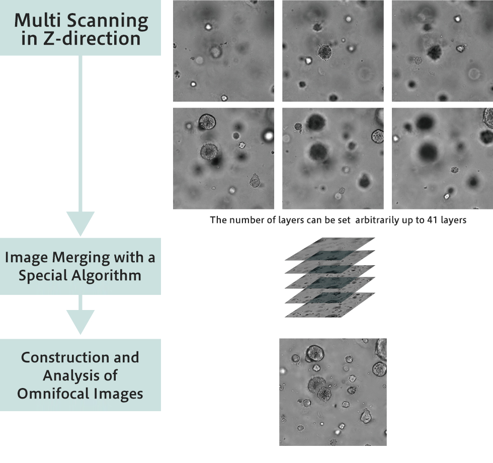
3D cellular imaging
- Equipped with a proprietary lens with a deep depth of field suitable for 3D cultured cell imaging and illumination
- Z-stacking imaging and focus composition functions
- Compatible with F-bottom, V-bottom, U-bottom, various SBS formats and microwell plates as standard
- Functional specialty plates such as plates for 3D culture, Corning® Elplasia® (Corning Japan) and EZSPHERE (AGC Techno Glass Co., Ltd.) are also available
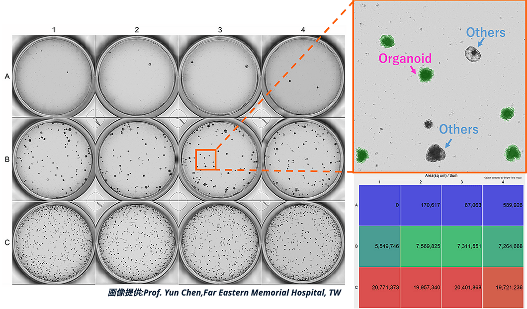
Deep Learning Plug-in (optional)
- Deep Learning to extract and quantify only grown organoids
- High confluence and non-uniform images can be accurately extracted and measured using Deep Learning
Multi-fluorescence imaging
- Multi-fluorescence imaging and analysis
- Bright field and 3 fluorescent colors* are automatically and
continuously imaged with the simultaneous loading of LED fluorescent filter * Cell3iMager duos2 only - Line up of 5 types of fluorescent filters
Automation system
- Automation by connecting external devices such as plate stacker and incubator with a plate transfer robot
- Automatically captures up to 200 images per day by robotization, streamlining a complicated workflow
Neurite elongation plug-in (optional)
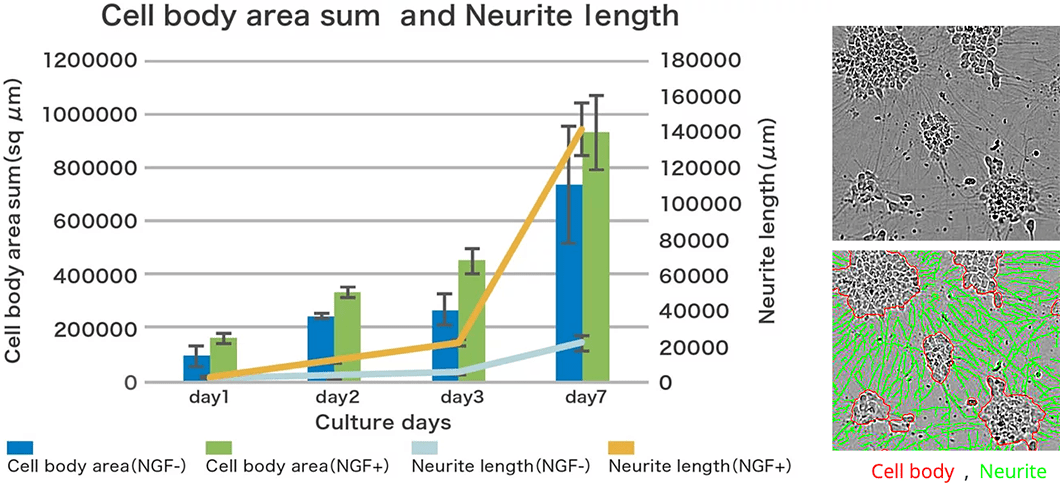
- Multi-object analysis, such as neurite elongation, is provided as a plug-in software
Publication
-
Novel bone microenvironment model of castration‑resistant prostate cancer with chitosan fiber matrix and osteoblasts
Masahiro Samoto Hideyasu Matsuyama Hiroaki Matsumoto Hiroshi Hirata Koji Ueno Sho Ozawa Junichi Mori Ryo Inoue Seiji Yano Yoshiaki Yamamoto Jun Haginaka Shizuyo Horiyama Koji Tamada
August 1, 2021 https://doi.org/10.3892/ol.2021.12950 Oncology Letters Article Number: 689 -
Mammary cell gene expression atlas links epithelial cell remodeling events to breast carcinogenesis
Kohei Saeki, Gregory Chang, Noriko Kanaya, Xiwei Wu, Jinhui Wang, Lauren Bernal, Desiree Ha, Susan L. Neuhausen, and Shiuan Chen
Commun Biol. 2021; 4: 660. Published online 2021 Jun 2. doi:10.1038/s42003-021-02201-2 -
Lactate Reprograms Energy and Lipid Metabolism in Glucose-Deprived Oxidative Glioma Stem Cells
Noriaki Minami ORCID,Kazuhiro Tanaka ,Takashi Sasayama ,Eiji Kohmura ORCID,Hideyuki Saya andOltea Sampetrean
Metabolites 2021, 11(5), 325; https://doi.org/10.3390/metabo11050325 Published: 18 May 2021
APPLICATION
-
Disease modeling and imaging assays using human iPS cell-derived airway organoids
Respiratory diseases include a wide range of conditions, including
chronic progressive diseases and acute and severe diseases such as
the recent COVID-19 pneumonia. However, the development of
fundamental therapeutics in this field has been slow, partly because
animal models such as mice used for preclinical drug efficacy evaluation
often do not reflect human disease states. A typical example is
cystic fibrosis (CF), an autosomal recessive genetic disorder caused
by mutations in the cystic fibrosis transmembrane conductance
regulator (CFTR), a chloride ion channel. -
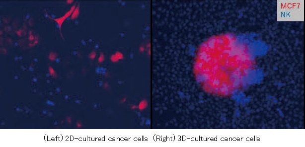
NK cell killing against 2D/3D-cultured cancer cells
In recent years, NK (Natural Killer) cells have been attracting attention in the fields of drug discovery development and cancer research. NK cells are a type of lymphocyte that works in innate immunity and play a major role in the removal of tumor cells and virus-infected cells.
By using Cell3iMager duos / duos2 and Deep Learning Plug-In, you will be able to perform a long-term Killing Assay. Since the imager uses a plate-fixed imaging method, clear imaging is possible even for suspended-cultured NK cells and 3D-cultured cancer cells. -
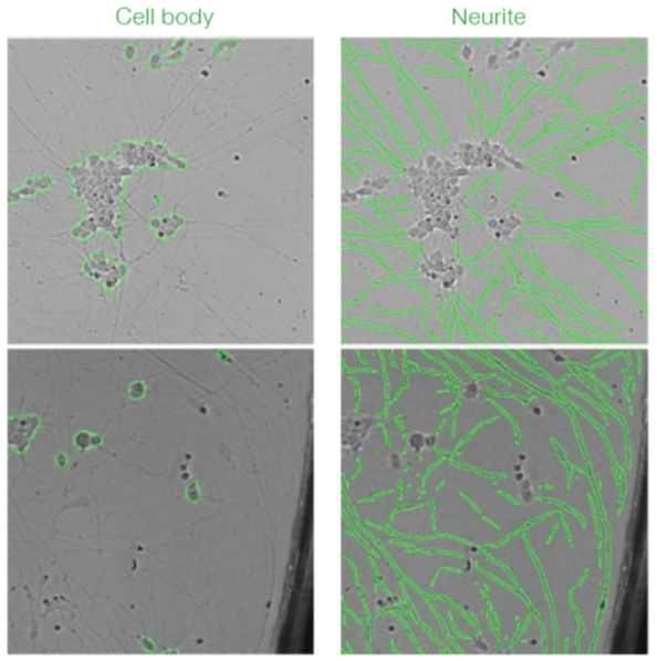
Imaging and Deep Learning based analysis of neurite outgrowth
With this study we conclude that Cell3iMager duos imaging technology in conjunction with deep learning is highly suited for delineating various biological processes using sympathetic neurons derived from hiPSC. Additionally, the platform can be used for identifying drug entities that could stimulate or block neuronal outgrowth. The analysis by deep learning accurately detects cells and is highly robust compared to traditional analyses even if there are differences in brightness or in the presence of debris.
-
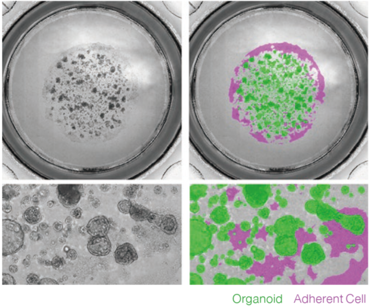
Label free imaging and analysis of organoids using Cell3iMager
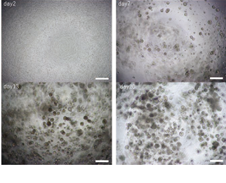
Cell3iMager duos / duos2 technology is highly suited for brightfield/fluorescence imaging of 3D cultured organoids. We conclude that the Label-free analysis is highly advantageous and could substitute microscopic method of monitoring of organoid growth processes, as segmentation of each organoid shape is possible using Deep Learning. Similarly, label-free analysis may also substitute ATP assays, toxicity assessments performed by fluorescent staining, and FIS assays.
-
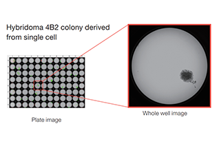
Single cell cloning using Hybridoma
Isolation of single cell derived clone with Duos superior imaging technology.
-
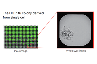
Single Cell Cloning
Utility of Cell3iMager duos imaging platform for isolation and assessment of single cell derived clone
-
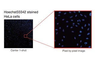
Counting fluorescence stained adherent cells
The cell density that stepwise cell number seeding, was quantyfied by using Cell3iMager duos.
-
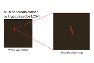
Evaluation of hypoxia level in 2D/3D-cultured cells
Hypoxia level of 2D/3D-cultured cells was evaluated by using Cell3iMager duos.
-
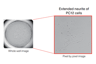
Neuronal Differentiation
Differentiation efficiency of β-NGF-treated PC12 was evaluated by using Cell3iMager duos.
-
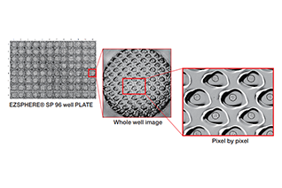
Analysis of spheroids in microwells (EZSPHERE® plate)
Spheroids were cultured with EZSPHERE® PLATE and analyzed using Deep Learning plug-in.
-
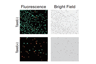
Live/Dead Assay
Cell viability in the presence of drug is calculated by using Calcein-AM/PI-double staining.
-
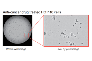
Cell Viability and Cytotoxicity Assay
Viability of anti-cancer drug treated cells is quantified by using Cell3iMager duos.
-
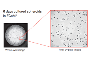
3D Cell Culture by using FCeM®
Multi spheroids cultured in FCeM-medium were quantified by using Cell3iMager duos.
-
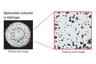
Assessing the growth of 3D spheroids cultured in Matrigel®
Visualisation and Quantification of spheroid volumes cultured in Cell3iMager duos.
-
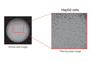
Confluency
The cell density that stepwise cell number seeding, is quantified by using Cell3iMager duos.
-
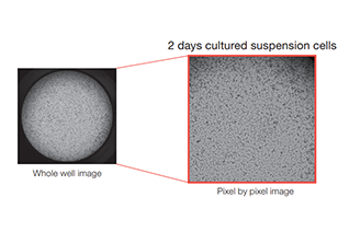
Suspension cultured cells
Suspension cultured cells were quantified by using Cell3iMager duos.
-
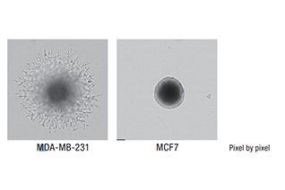
3D Invasion Assay
Invasiveness of MCF-7 (low malignant tumor) and MDA-MB-231 (high malignant tumor ) cells are compared by using Cell3iMager duos.
-
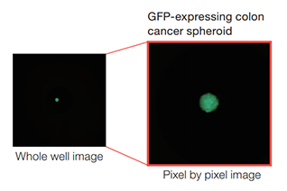
Anti-Cancer Drug Screening
Anti-cancer drug screening in TCF4/LEF reporter (GFP) -expressing cells is performed by using Cell3iMager duos.
-
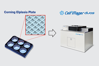
Label free analysis of spheroids in microwells (Corning® Elplasia® Plates)
Spheroids in microwells of Corning®Elplasia® plate made by Corning are scanned and measured by Cell3iMager duos.
-
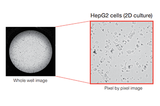
Cell Proliferation
Proliferation ratio of the 2D/3D cultured cells is quantified by Cell3iMager duos.
-
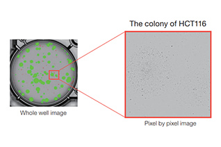
Label free colony-forming units assay
We performed the labeled-free colony formation assay using Cell3iMager duos.
-
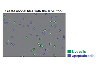
Deep Learning for cell classification
Jarkat cells induced cell death by Anti-Fas antibody were classified by Cell3iMager duos and Deep Learning.
-
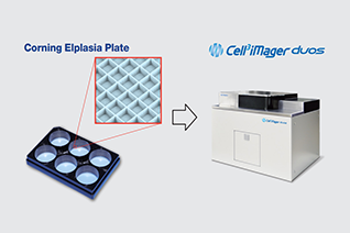
Label free analysis of spheroids in microwells (Corning® Elplasia® Plates)
Spheroids in microwells of Corning®Elplasia® plate made by Corning are scanned and measured by Cell3iMager duos.
Publication
-
Artificial intelligence-based analysis of time-lapse images of sphere formation and process of plate adhesion and spread of pancreatic cancer cells
Yuuki Shichi, Fujiya Gomi1, Yasuko Hasegawa, Keisuke Nonaka, Seiichi Shinji, Kimimasa Takahashi, Toshiyuki Ishiwata
-
Modeling chemotherapy induced neurotoxicity with human induced pluripotent stem cell (iPSC) -derived sensory neurons
Christian Schinke, Valeria Fernandez Vallone , Andranik Ivanov , Yangfan Peng , Péter Körtvelyessy , Luca Nolte , Petra Huehnchen , Dieter Beule , Harald Stachelscheid , Wolfgang Boehmerle , Matthias Endres
SPECIFICATIONS
| Product name (Code) | Cell3iMager duos2(CC-8300) | Cell3iMager duos(CC-8000) |
|---|---|---|
| Channel | Bright field and 3 Colors of Fluorescence | Bright field and 1 Colors of Fluorescence |
| Bright field light source | White LED strobes | White LED strobes |
| Camera | CMOS 4.2 megapixel color | CMOS 4.2 megapixel color |
| Lenses | Original hypercentric lens (High-speed mode) Original telecentric lens (High-quality mode) |
Original hypercentric lens (High-speed mode) Original telecentric lens (High-quality mode) |
| Resolution | 4.0μm/pixel (High-speed mode) 0.8μm/pixel (High-quality mode) |
4.0μm/pixel (High-speed mode) / 0.8μm/pixel (High-quality mode) |
| Auto focus | HW: Laser type real time auto focus SW: Image contrast software auto focus |
HW: Laser type real time auto focus SW: Image contrast software auto focus |
| Image output | 24bit color (8bit×3) | 24bit color( 8bit×3) |
| Fluorescent filter type | DAPI, GFP, Cy3, Texas Red, Cy5 | - |
| Fluorescent light source (excitation wavelength) | - | U : 384nm , B : 470nm , G : 530nm , Y565nm , R : 625nm |
| Internal temperature | 35℃±2℃ automatic adjustment, during the power is on | 35℃±2℃ automatic adjustment, during the power is on (optional) |
| Placement environment | temperature 18-28℃, humidity 80% or less, no condensation | temperature 18-28℃, humidity 80% or less, no condensation |
| Transport conditions | Packaged: 0-55℃, humidity 80% or less, no condensation | Packaged: 0-55℃, humidity 80% or less, no condensation |
| Culture container | 6・12・24・48・96・384 microwell plate (Compatible with almost all SBS standard plates) 35・60・100mm dish, slide glass (Optional adapter required) |
6・12・24・48・96・384 microwell plate (Compatible with almost all SBS standard plates) 35・60・100mm dish, slide glass (Optional adapter required) |
| Power supply | AC100-240V / 250VA | AC100-240V / 250VA |
| Size and Weight | W677xD580xH550 mm / 111 kg | W677 x D570 x H550 mm / 110kg |
| Software | Dedicated Cell3iMager software, includes as standard | Dedicated Cell3iMager software, includes as standard |
| Designated computer with confirmed operation | HP Z4 G4 workstation( SCREEN designated configuration), OS: Windows10 | HP Z4 G4 workstation( SCREEN designated configuration), OS: Windows10 |
| Product name (Code) | Channel | Bright field light source | Camera | Lenses | Resolution | Auto focus | Image output | Fluorescent filter type | Fluorescent light source (excitation wavelength) | Internal temperature | Placement environment | Transport conditions | Culture container | Power supply | Size and Weight | Software | Designated computer with confirmed operation |
|---|---|---|---|---|---|---|---|---|---|---|---|---|---|---|---|---|---|
| Cell3iMager duos2 (CC-8300) |
Bright field and 3 Colors of Fluorescence | White LED strobes | CMOS 4.2 megapixel color | Original hypercentric lens (High-speed mode) Original telecentric lens (High-quality mode) |
4.0μm/pixel (High-speed mode) 0.8μm/pixel (High-quality mode) |
HW: Laser type real time auto focus SW: Image contrast software auto focus |
24bit color (8bit×3) | DAPI, GFP, Cy3, Texas Red, Cy5 | - | 35℃±2℃ automatic adjustment, during the power is on | temperature 18-28℃, humidity 80% or less, no condensation | Packaged: 0-55℃, humidity 80% or less, no condensation | 6・12・24・48・96・384 microwell plate (Compatible with almost all SBS standard plates) 35・60・100mm dish, slide glass (Optional adapter required) |
AC100-240V / 250VA | W677 x D580 x H550 mm / 111kg | Dedicated Cell3iMager software, includes as standard | HP Z4 G4 workstation( SCREEN designated configuration), OS: Windows10 |
| Cell3iMager duos (CC-8000) |
- | U : 384nm , B : 470nm , G : 530nm , Y565nm , R : 625nm |
35℃±2℃ automatic adjustment, during the power is on (optional) | W677 x D570 x H550 mm / 110kg |