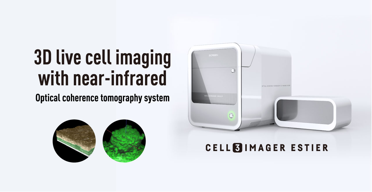
PRODUCT
3D morphology and internal structure
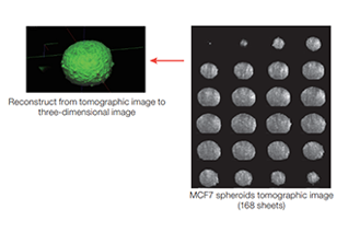
- OCT is the technique that can acquire the tomogram of the live cell or organism without aggression .Detailed observation for 3D structure of spheroids that conventional microscopes cannot recognize
Noninvasive and timelapse
- Non-invasive, real-time observation of tomographic images and 3D morphology of spheroids, from their formation to collapse after addition of anti-cancer drugs, using OCT technology with weak near-infrared light
Easy operation
- Simple operation on the PC screen.
- Unnecessary of special training & expert technique.
- Compatible with general micro well plates (SBS format) and culture dish.Because of the same workflow with microscope, it enables to reduce experiment cost and burden on researcher
FEATURES
3D morphological analyses for cultures / internal structures with OCT
OCT is the technique that can acquire the tomogram of the live cell or organism without aggression by using feeble near-infrared light.
Researchers can acquire 3D image safety and in real time without having any obstacles action for the living body as they use our device.
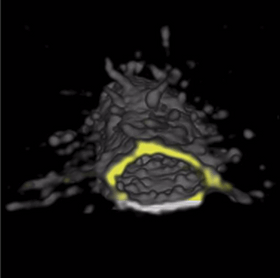
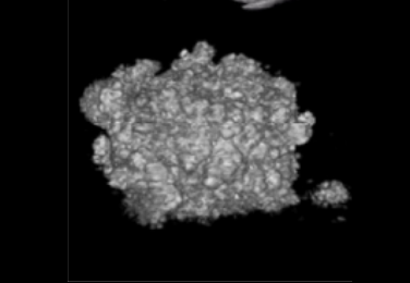
Observation of internal structure
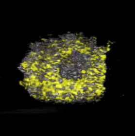
- Spheroid internal cavity morphology(Prof. Tamio Mizukami, Nagahama Institute of Bio-Science and Technology)
- Neovasculature(Prof. Yukiko T. Matsunaga, University of Tokyo)
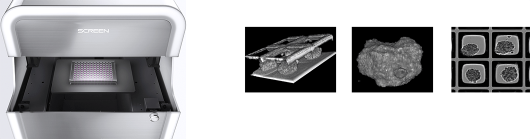
Tomographic imaging of living organisms/cell microstructure imaging
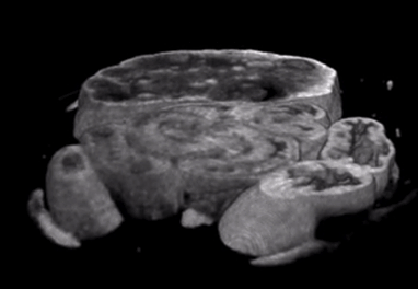
- Ovary(Prof. Nobuo Nagai, Nagahama Institute of Bio-Science and Technology)
- Kidney spheroid(Prof. Tetsuya Ohbayashi, Tottori University)
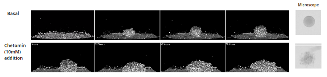
Time-lapse observation by non-invasive and label-free analysis
- Real time observation from spheroid formation to collapse after addition of anti-cancer drug
- Detailed observation for 3D structure of spheroids that conventional microscopes cannot recognize
Compatibility for conventional labwares
- Compatible with general micro well plates (SBS format) and culture dish
- Specific well plates optimized for 3D culture such as "Corning® Elplasia® " is also available
Publication
-
Development of a 3D Cell Culture System Using Amphiphilic Polydepsipeptides and Its Application to Hepatic Differentiation
Junko Enomoto, Yukiko Toba, Haruka Yamazaki, Masaki Kanai, Hiroyuki Mizuguchi, and Hayato Matsui
ACS Appl. Bio Mater. 2021, 4, 9, 7290–7299 Publication Date:September 8, 2021 https://doi.org/10.1021/acsabm.1c00816 -
Development, Characterization and Potential Applications of a Multicellular Spheroidal Human Blood–Brain Barrier Model Integrating Three Conditionally Immortalized Cell Lines
Keita Kitamura, Kenta Umehara, Ryo Ito, Yoshiyuki Yamaura, Takafumi Komori, Hanae Morio, Hidetaka Akita, Tomomi Furihata
Biological and Pharmaceutical Bulletin 2021 Volume 44 Issue 7 Pages 984-991 DOI https://doi.org/10.1248/bpb.b21-00218 -
Three-Dimensional Live Imaging of Bovine Preimplantation Embryos: A New Method for IVF Embryo Evaluation
Yasumitsu Masuda, Ryo Hasebe, Yasushi Kuromi, Masayoshi Kobayashi, Kanako Urataki, Mitsugu Hishinuma, Tetsuya Ohbayashi and Ryo Nishimura
Veterinary Reproductive Immunology Front. Vet. Sci., 26 April 2021 | https://doi.org/10.3389/fvets.2021.639249
APPLICATION
-
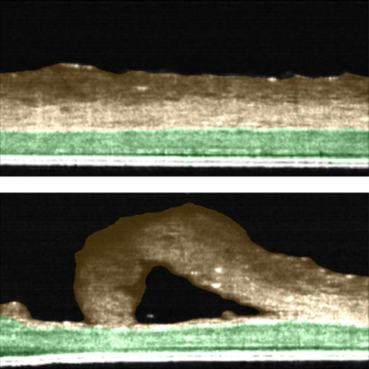
Skin Irritation test : Evaluation of thickness in skin model using OCT technology
To summarize, we observed that by using the low magnification mode, we were able to obtain the thickness information of the entire 24-well insert. In high magnification mode, we were able to obtain thickness information of two layers, stratum corneum and cell layer, non-invasively. We conclude that the Cell3iMager Estier equipped with OCT technology can be used to measure the thickness of stacked cell sheets in real time and implemented as “Skin irritation test” to evaluate safety of topical creams/agents developed by Biotech and Pharma industry.
-
Thickness of cell sheet
NHDF sheets were imaged by Cell3iMager Estier, and thickness of sheets were calculated from these images.
-
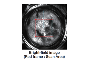
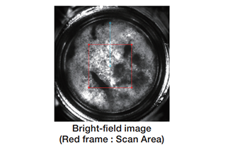
Real volume of spheroids
The volume of spheroids that stepwise cell number seeding, is quantified by using Cell3iMager Estier.
-
Thickness of cell sheet
NHDF sheets were imaged by Cell3iMager Estier, and thickness of sheets were calculated from these images.
-

Real volume of spheroids
The volume of spheroids that stepwise cell number seeding, is quantified by using Cell3iMager Estier.
Publication
-
In-process monitoring of a tissue-engineered oral mucosa fabricated on a micropatterned collagen scaffold: use of optical coherence tomography for quality control
O. Suebsamarn , Y. Kamimura , A. Suzuki , Y. Kodama , R. Mizuno , Y. Osawa , T. Komatsu , T. Sato , K. Haga , R. Kobayashi , E. Naito , M. Kida , K. Kishimoto , J. Mizuno k , H. Hayasaki , K. Izumi
-
Scalable production of homogeneous cardiac organoids derived from human pluripotent stem cells
Taijun Moriwaki Hidenori Tani Kotaro Haga Yuika Morita-Umei Yusuke Soma Tomohiko C. Umei Otoya Sekine Kaworu Takatsuna Yoshikazu Kishino Hideaki Kanazawa Jun Fujita Keiichi Fukuda Shugo Tohyama Masaki Ieda
-
Multiple cystic sphere formation from PK-8 cells in three-dimensional culture
YuukiShichi FujiyaGomi YoshibumiUeda KeisukeNonaka FumioHasegawa YasukoHasegawa NaeHinata HisashiYoshimura MasamiYamamoto KimimasaTakahashi TomioArai ToshiyukiIshiwa
SPECIFICATIONS
| Product name (Code) | Cell3iMager Estier (CC-9000) |
|---|---|
| Data | Tomogram in user indicated location / 3D image from user indicated view point / Movie output of tomogram / Animation output of 3D image |
| Output parameters | Quantified value: distance between point to point, area of 2D image , volume, sphericity, surface rough degree, cavity volume |
| Light Source | Near-infrared SLD light source (OCT type) |
| Max. FOV | High resolution: 1 x 1 mm, Low resolution: 10 x 10 mm (Wide F.O.V.) |
| Max. depth | 1,000um |
| Resolution | High resolution: 3 um, Low resolution: 10 um |
| Cross-sectional observation time (e.g.) | 0.5 sec. or more |
| 3D observation time (e.g.) | 3D observation: High resolution 0.3 x 0.3 x 0.3 mm / 3 μm: 1 min. / Low resolution 5.0 x 5.0 x 1.0 mm / 10 μm: 9 min. |
| Placement environment | temperature 18-28℃, humidity 80% or less, no condensation |
| Transport conditions | Packaged: 0-55℃, humidity 80% or less, no condensation |
| Culture container | 6・12・24・48・96・384 microwell plate (Compatible with almost all SBS standard plates) / 35・60・100mm dish, slide glass (Optional adapter required) |
| Incubator | Temperature (35~42°C), CO2 (Optional) |
| Power supply | AC100-240V / 200VA |
| Size and Weight | Main unit:W420 x D530 x H500 mm / 50kg Sub unit :W406 x D230 x H190 mm / 7kg |
| Software | Dedicated Cell3iMager software, includes as standard |
| Designated computer with confirmed operation | HP Z4 G4 workstation( SCREEN designated configuration), OS: Windows10 |
| Product name (Code) | Data | Output parameters | Light Source | Max. FOV | Max. depth | Resolution | Cross-sectional observation time (e.g.) | 3D observation time (e.g.) | Placement environment | Transport conditions | Culture container | Incubator | Power supply | Size and Weight | Software | Designated computer with confirmed operation |
|---|---|---|---|---|---|---|---|---|---|---|---|---|---|---|---|---|
| Cell3iMager Estier (CC-9000) |
Tomogram in user indicated location / 3D image from user indicated view point / Movie output of tomogram / Animation output of 3D image |
Quantified value: distance between point to point, area of 2D image , volume, sphericity, surface rough degree, cavity volume |
Near-infrared SLD light source (OCT type) | High resolution: 1 x 1 mm, Low resolution: 10 x 10 mm (Wide F.O.V.) |
1,000um | High resolution: 3 um, Low resolution: 10 um |
0.5 sec. or more | 3D observation: High resolution 0.3 x 0.3 x 0.3 mm / 3 μm: 1 min. / Low resolution 5.0 x 5.0 x 1.0 mm / 10 μm: 9 min. |
temperature 18-28℃, humidity 80% or less, no condensation |
Packaged: 0-55℃, humidity 80% or less, no condensation |
6・12・24・48・96・384 microwell plate (Compatible with almost all SBS standard plates) / 35・60・100mm dish, slide glass (Optional adapter required) |
Temperature (35~42°C), CO2 (Optional) |
AC100-240V / 200VA | Main unit:W420 x D530 x H500 mm / 50kg Sub unit :W406 x D230 x H190 mm / 7kg |
Dedicated Cell3iMager software, includes as standard |
HP Z4 G4 workstation (SCREEN designated configuration), OS: Windows10 |