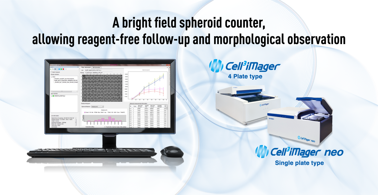

PRODUCT
Quantification of 3D cultured cells in just 90 seconds
- Bright-field, label-free, non-invasive measurement
- Automatic imaging and quantification at a reasonable price
High-throughput imaging

Label-free analysis enables continuous observation
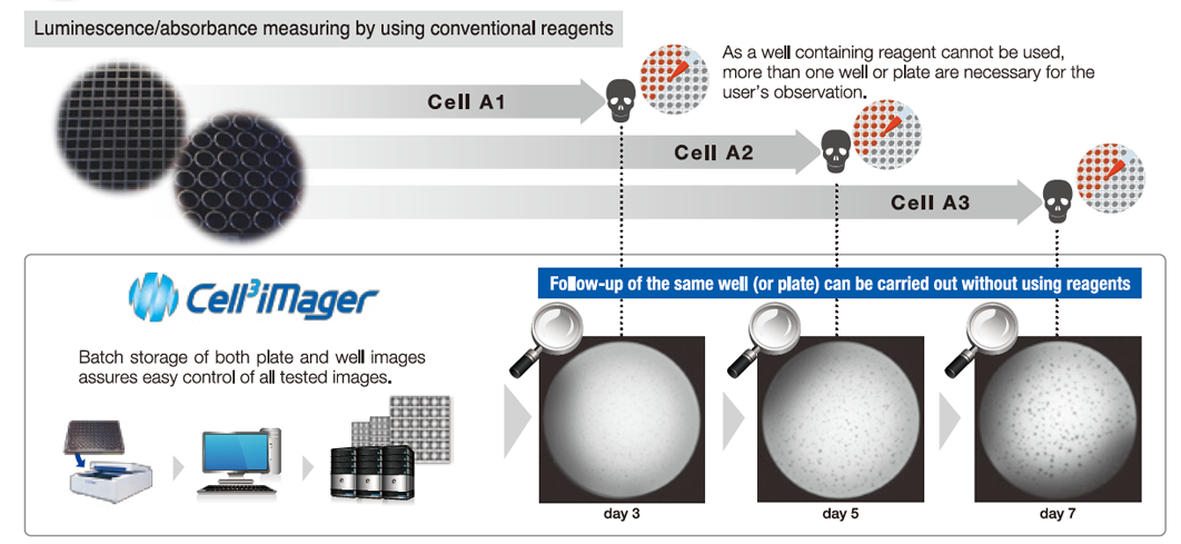
Label-free and non-invasive analysis using bright field, allowing automatic measurement of course and morphology in the same wells, and continuous observation to graph the course of growth and save images.
Affordable and Automated Imaging
The Entry Model to High-Speed Imaging System, specialized for 3D cultured cells, provides the convenience of automated imaging and analysis at an affordable price.
FEATURES
High-throughput imaging
- Rapid quantification of 96 wells in 90 seconds
- High speed imaging and high speed analysis make 3D cultured cell quantification more accurate and efficient.
- In addition, it supports automation, allowing users to build a workflow that suits them, such as scanning first and analysis later.

Imaging system specialized for 3D culture
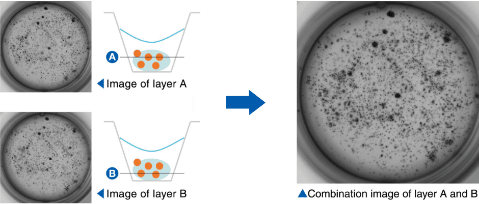
- All-in-focus images, which are synthesized from images captured at multiple depths of focus, can be used to analyze cells scattered in the thickness direction.
- Integrating information in the thickness direction allows accurate quantification of three-dimensional cultures, such as agar and floating culture systems.
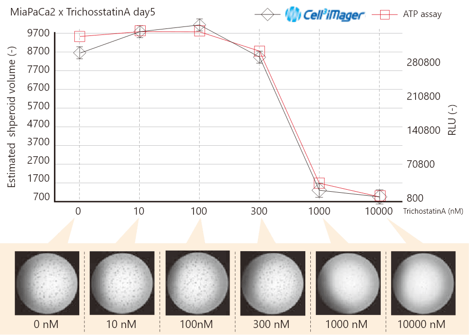
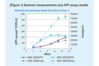
High correlation with ATP assay
- Software and transillumination provide accurate supplementation to spheroids around the wells and analogize the volume value of spheroids.
Time course statistics
- Different culture environments and compound test patterns can be grouped in a well plate.
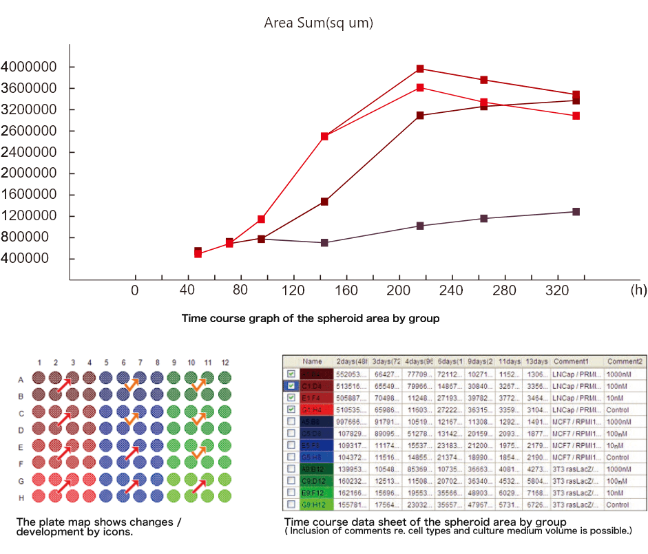
Selectable scan resolution
- Adjustable scanning accuracy from 200 dpi to 9600 dpi (2.6 μm/pixel) to match the sample and analysis speed(7 levels: 200, 300, 600, 1200, 2400, 4800, 9600 dpi)
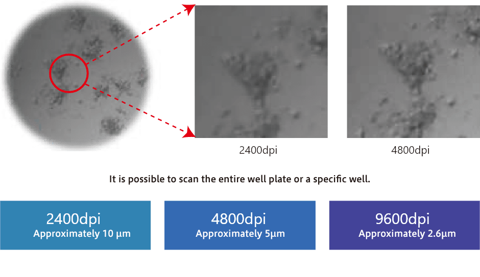
Software for 3D Culture
- Multi-focus scanning in the Z-axis ensures that no cells are missed, even in 3D cultures such as gel-embedded cultures.
- Supports a variety of analytical screening such as kinetic analysis and temporal analysis.
- More than 30 parameters are employed to accurately separate the sample signal from the artificial signal.
- This enables accurate extraction and viability determination of cultured cells.
- Supports quantification of single spheroids and multiple spheroids.
Publication
-
Gamma-Mangostin Isolated from Garcinia mangostana Suppresses Colon Carcinogenesis and Stemness by Downregulating the GSK3β/β-Catenin/CDK6 Cancer Stem Pathway
Alexander THWu ,Yuan-ChiehYeh ,Yan-JunHuang ,NtlotlangMokgautsi ,BashirLawal ,Tse-HungHuang
Phytomedicine Available online 21 October 2021, 153797 DOI:https://doi.org/10.1016/j.phymed.2021.153797 -
speedingCARs: accelerating the engineering of CAR T cells by signaling domain shuffling and single-cell sequencing
Raphaël B. Di Roberto, ProfileRocío Castellanos-Rueda, Fabrice S. Schlatter, Darya Palianina, ProfileOanh T. P. Nguyen, Edo Kapetanovic, ProfileAndreas Hierlemann, ProfileNina Khanna, ProfileSai T. Reddy
biorxiv Posted August 23, 2021. doi: https://doi.org/10.1101/2021.08.23.457342 -
Hypoxia-targeted cupric-tirapazamine liposomes potentiate radiotherapy in prostate cancer spheroids
Vera L.Silva, AmaliaRuiz ,AhlamAli ,SaraPereira ,JaniSeitsonen ,JanneRuokolainen ,FionaFurlong ,JonathanCoulter Wafa' T.Al-Jamal
International Journal of PharmaceuticsVolume 607, 25 September 2021, 121018 doi:https://doi.org/10.1016/j.ijpharm.2021.121018 -
Differential Angiogenic Potential of 3-Dimension Spheroid of HNSCC Cells in Mouse Xenograft
So-Young Choi ,Soo Hyun Kang ,Su Young Oh ,Kah Young Lee ,Heon-Jin Lee ,Sangil Gum ,Tae-Geon Kwon ,Jin-Wook Kim ,Sung-Tak Lee ,Yoo Jin Hong ,Dae-Geon Kim and Su-Hyung Hong
Int. J. Mol. Sci. 2021, 22(15), 8245; https://doi.org/10.3390/ijms22158245 Published: 31 July 2021
APPLICATION
-
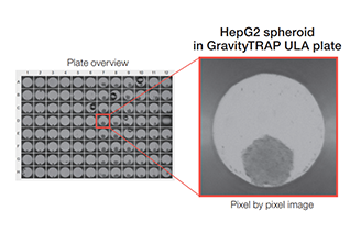
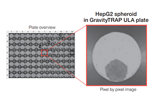
3D Cell Culture in Gravity TRAP™
Spheroids formed by the method of hanging drop were moved to Gravity TRAPTM ULA plate, and quantified by Cell3iMager neo.
-
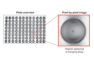
3D Cell Culture by Hanging Drop
Spheroids in the droplets being aggregated by the method of hanging drop were quantified by Cell3iMager neo
-
Application note Vol.1
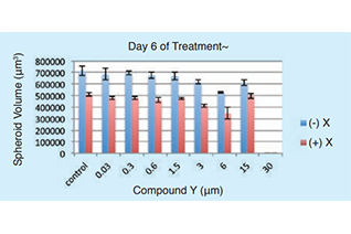
Development of spheroid-based anticancer drug screening method using high-speed cell culture scanner
-
Application note Vol.3
High-Speed 3D Cell Scanner —Perfect for the Study of Complex 3D MCTS Models
-
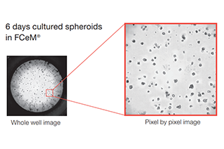
3D cell culture by using hydrogels
Spheroids cultured in hydrogel were quantified by using Cell3iMager neo.
-
3D Cell Culture in Gravity TRAP™
Spheroids formed by the method of hanging drop were moved to Gravity TRAPTM ULA plate, and quantified by Cell3iMager neo.
-
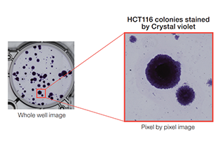
Colony-forming units assay
Cell3iMager neo is useful for ‘colony formation assay’ stained by crystal violet
-
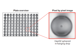
3D Cell Culture by Hanging Drop
Spheroids in the droplets being aggregated by the method of hanging drop were quantified by Cell3iMager neo.
-
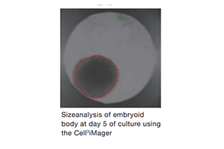
Rapid production and size assessment of Embryoid Bodies ( EBs )
Simplify formation of EBs with GravityPLUS™ hanging drop plates and Cell3 iMager analysis
-
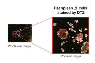
Pancreatic β cells stained by DTZ
Pancreatic β cells isolated from Rat spleen was quantified by Cell3iMager neo. To detect only β cells, isolated cells were stained by DTZ.
-

3D Cell Culture in Gravity TRAP™
Spheroids formed by the method of hanging drop were moved to Gravity TRAPTM ULA plate, and quantified by Cell3iMager neo.
SPECIFICATIONS
| Product name (Code) | Cell3iMager (CC-5000) | Cell3iMager neo (CC-3000) |
|---|---|---|
| Channel | Bright field | |
| Light source | Wite LED transmission illuminations | |
| Camera | CCD line sensor (4096 elements) | |
| Lenses | Original low-depth-of-field lens | |
| Resolution | 200dpi,300dpi,600dpi,1200dpi,2400dpi,4800dpi,9600dpi | |
| Auto focus | Image contrast software auto focus | |
| Image output | 8bit mono | |
| Placement environment | temperature 18-28℃, humidity 80% or less, no condensation | |
| Transport conditions | Packaged: 0-55℃, humidity 80% or less, no condensation | |
| Culture container | 6・12・24・48・96・384 microwell plate (Compatible with almost all SBS standard plates) / 35・60mm dish, slide glass (Optional adapter required) |
6・12・24・48・96・384 microwell plate (Compatible with almost all SBS standard plates) / 35・60・100mm dish, slide glass (Optional adapter required) |
| Power supply | AC100-240V / 250VA | |
| Size and Weight | W492 x D775 x H294 mm / 40kg | W594 x D381 x H294 mm / 25kg |
| Software | Dedicated Cell3iMager software, includes as standard | |
| Plate number | 4 | 1 |
| Designated computer with confirmed operation | HP Elite Desk 800 , OS: Windows10 | |
| Product name (Code) | Channel | Light source | Camera | Lenses | Resolution | Auto focus | Image output | Placement environment | Transport conditions | Culture container | Power supply | Size and Weight | Software | Plate number | Designated computer with confirmed operation |
|---|---|---|---|---|---|---|---|---|---|---|---|---|---|---|---|
| Cell3iMager (CC-5000) | Bright field | Wite LED transmission illuminations | CCD line sensor (4096 elements) | Original low-depth-of-field lens | 200dpi,300dpi,600dpi,1200dpi,2400dpi,4800dpi,9600dpi | Image contrast software auto focus | 8bit mono | temperature 18-28℃, humidity 80% or less, no condensation | Packaged: 0-55℃, humidity 80% or less, no condensation | 6・12・24・48・96・384 microwell plate (Compatible with almost all SBS standard plates) / 35・60mm dish, slide glass (Optional adapter required) |
AC100-240V / 250VA | W492 x D775 x H294 mm / 約40kg | Dedicated Cell3iMager software, includes as standard | 4 | HP Elite Desk 800 , OS: Windows10 |
| Cell3iMager neo (CC-3000) | 6・12・24・48・96・384 microwell plate (Compatible with almost all SBS standard plates) / 35・60・100mm dish, 25cm2 flask, slide glass (Optional adapter required) |
W594 x D381 x H294 mm / 25kg | 1 |